Avascular Necrosis of the Hip (AVN)
Authors
Dr Keran Sundaraj MBBS, MSc (Trauma), FRACS, FAOA
Avascular Necrosis of the Hip (AVN)
Avascular necrosis (AVN or osteonecrosis) of the hip is a painful condition that is caused by a disruption of the blood supply to the femoral head (top of the thigh bone).
Anatomy
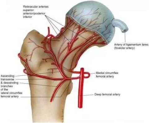
The hip is a ball and socket joint. The socket is formed by the acetabulum (part of the pelvis), and the ball is formed by the femoral head (upper end of the thigh bone). The blood supply arises from three parts;
The capsular (lining of the joint)
The bone
Ligament of Teres - inside the joint
This blood supply is unusual as the main blood supply runs from below up. This makes it more difficult for blood to arrive at the femoral head. Thus a minor disruption can significantly change the amount of blood the femoral head receives.
Cause
Primary or secondary pathologies may cause disruption in the blood supply to the femoral head:
Primary
Idiopathic (meaning unknown cause)
Secondary
Excess alcohol intact
Injuries - such as fracture or dislocation
Steroids - in high dose and prolonged use. Generally, short term 'pulse' therapy does not cause AVN.
Medical conditions - blood disorders or diver's disease ("the bends")
Eventually, the bone dies off due to the lack of blood flow. This causes the joint end to loose its protective cartilage cap, and the joint surface becomes irregular. The surfaces are no longer smooth and free running, and this leads to stiffness and pain. Eventually, the joint wears away to such an extent that the bone upper end of the femur rubs on the acetabulum. This leads to arthritis.
Imaging Tests
X-rays
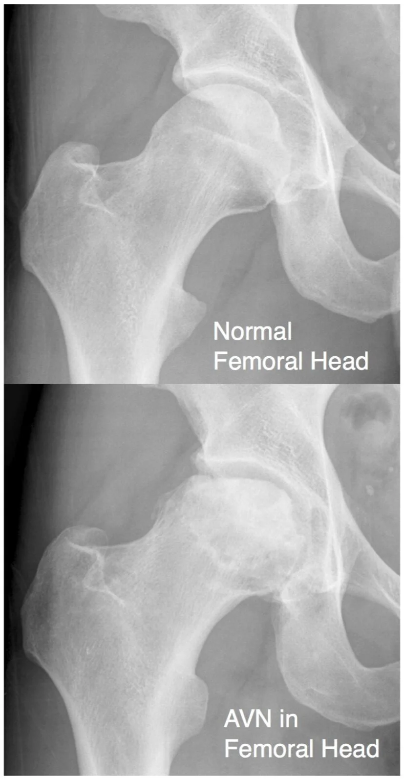
X-ray is used to assess the joint for arthritis and the severity of AVN. Findings on the X-ray do not necessarily correlate with the severity of a patients symptoms.
MRI
MRI is used in the early stages of the disease, before the femoral head surface becomes irregular. Areas of focal necrosis and cartilage thickness can be assessed with an MRI.
Bone Scan
A bone scan is helpful to assess blood flow to the femoral head. However, this is an older technique that the use of MRI has primarily surpassed.
Sometimes patients present with a bone scan that was ordered for a different reason showing AVN. This is known as an incidental finding.
Treatment
Non-surgical management
For patients that are not symptomatic, regular observation may be all that is required.
Treatment of the underlying cause (if there is one) will take place. However, given the nature of the diseases that cause AVN, there may not be much scope to change the underlying issue.
Core decompression
This technique uses drill holes to relieve pressure and create new blood flow channels into the necrotic area.
The results of this area variable, though if the AVN area is small and intervention is performed early enough, there is some chance of success.
Total hip replacement
If the AVN has progressed to cause the bone to collapse, then the most successful treatment is with a total hip replacement.
See the patient information sheet on total hip replacement for further information.
Get in touch.
Fill out the form and one of the team will be back in touch within 24 hours.
Alternatively, give us a call on
(02) 9437 5999


